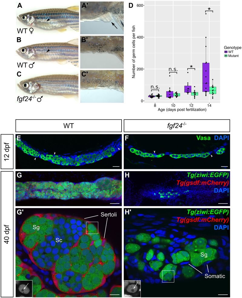
Fig 2. Larval and juvenile fgf24 mutants have severely underdeveloped gonads and consequently develop as male.
(A-C’) fgf24 mutants are phenotypically male. A wild-type (WT) female has a distended belly (A) and protruding cloaca (arrow, A’) compared to the slender belly and obscured cloaca of the wild-type (B, B’) and mutant (C, C’) males. Note the absence of pectoral fin in the fgf24 mutant (C) compared to wild-type animals (arrowheads, A, B). (D) Box and whisker plot representing the number of germ cells per wild-type and fgf24 mutant fish during larval development. Each dot represents the number of germ cells in one animal (Unpaired two-tailed t-test; n = 15 for both 8 dpf wild-type and mutant, n = 13 for 10 dpf wild-type, n = 12 for 10 dpf mutant, n = 10 for 12 dpf wild-type, n = 11 for 12 dpf mutant, n = 16 for 14 dpf wild-type, n = 12 for 14 dpf mutant; * = P < .05; n.s. = no significance). (E-H’) Representative confocal images of 12 dpf (E-F) and 40 dpf (G-H’) wild-type (E, G-G’) and fgf24 mutant (F, H-H’) gonads. At 12 dpf, gonads of fgf24 mutants (F) have fewer Vasa positive germ cells (green) compared to wild-type (E). By 40 dpf, wild-type testes have many Tg(ziwi:EGFP) positive germ cells (green), both premeiotic and spermatogenic, that are enclosed by Tg(gsdf:mCherry) positive Sertoli cells (red) (G-G’). In contrast, gonads of 40 dpf fgf24 mutants are unorganized, lack Tg(gsdf:mCherry) expressing Sertoli cells, and contain few germ cells, all of which are premeiotic (H-H’). G’ and H’ are magnified views G and H, respectively. The boxed nuclei in G’ and H’ are magnified in the respective insets, with an arrow indicating the large nucleoli (DAPI only, in grey). E-H’ are sagittal optical sections with anterior to the left. Nuclei labeled with DAPI (blue). Scale bars = 20 μm for E, F, G, H; 100 μm for G’, H’.

