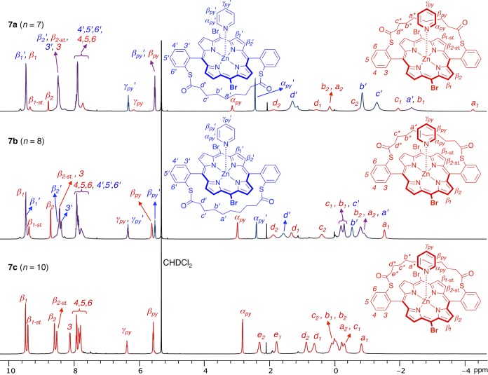
Figure 6.
1H NMR spectra of 1:1 complexes of strapped dibromoporphyrins 7a–c with pyridine in CD2Cl2 at 193 K (500 MHz). Signals belonging to the out and in complexes are displayed in blue and red, respectively, with overlapping signals displayed in purple. In the in complex, the reduced symmetry of the molecule leads to splitting of the β pyrrole proton signals into 4 doublets (β1-st and β2-st refer to the β protons on the strap side), and restricted conformational freedom leads to splitting of the strap proton signals (noted for example a1 and a2 for the a protons).

