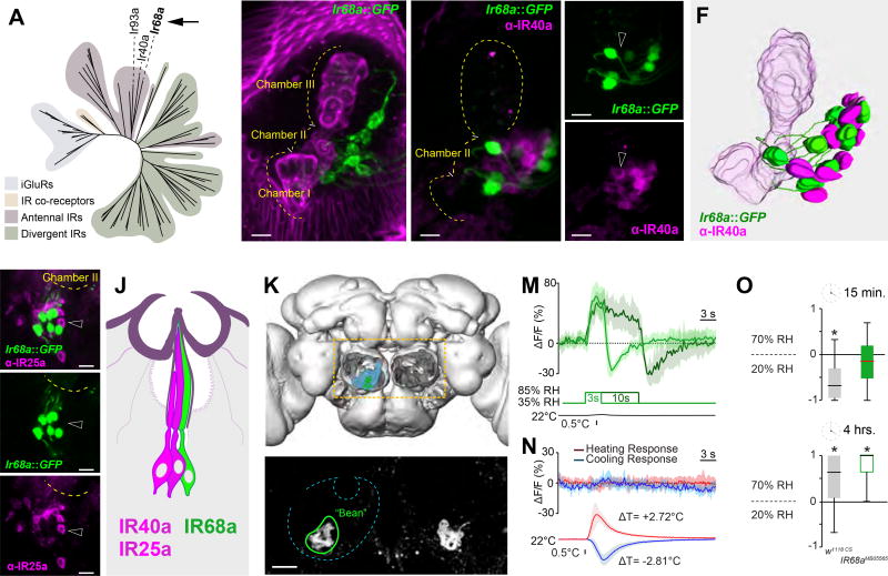
Figure 1. IR68a is expressed in sacculus neurons which mediate humid-air responses.
(A) Phylogenetic tree of the IR family, highlighting the position of IR68a.
(B) Ir68a-expressing neurons innervate chamber II of the sacculus, a small invagination of the antenna (Magenta= maximum-intensity projection of cuticular autofluorescence showing the sacculus; green= Ir68a>GFP neurons).
(C–E) Immunostaining of an Ir68a>GFP antenna using anti-IR40a antibodies (max projection of a shallow confocal stack, scalebar=10µm, dotted line=outline of the sacculus, arrowheads point to adjacent cells to highlight no overlap in expression).
(F) 3D model of the sacculus based on (C).
(G–I) Immunostaining of an Ir68a>GFP antenna using anti-IR25a antibodies (max projection of a shallow confocal stack, scalebar=10µm, dotted line=outline of the sacculus, arrowheads point to adjacent cells to highlight no overlap in expression).
(J) Cartoon of a hygrosensory sensillum.
(K) 3D reconstruction of the Drosophila brain, antennal lobes removed, to highlight the posterior antennal lobe (PAL, light blue).
(L) 2-photon stack of an Ir68a>GFP brain shows axonal termini converging into two symmetric, ‘bean’ shaped glomeruli (green outline) in the PAL (scalebar=10µm).
(M–N) Average traces from ‘bean’ in response to humid air (M) and temperature (N) stimuli (shaded areas represent STD).
(O) Humidity preference index of w1118 controls and Ir68aMB05565 mutants (see Figure S1 for additional timepoints). The edges of the boxes are the first and third quartiles, thick lines mark the medians, whiskers represent data range. Preference was tested with one-sample t test, theoretical mean 0, p < 0.05 was considered significant, i.e. no asterisk indicate no significant preference.

