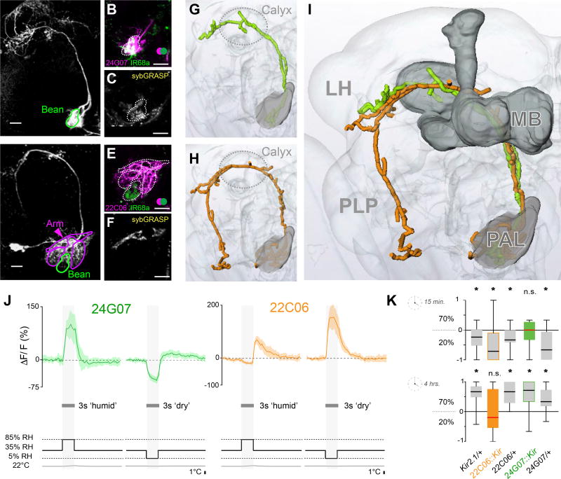
Figure 3. Projection neurons innervating hygrosensory glomeruli of the posterior antennal lobe.
(A–F) Maximum-intensity projections of two-photon imaging stacks of live brains expressing transgenic labels (scale bars =10µm). (A) R24G07> CD8:GFP labels hygrosensory PNs innervating the Bean (Se also Figure S2a). (B) R24G07> TdTomato; IR68a> CD8:GFP demonstates significant spatial overlap (green=CD8:GFP, pink=TdTomato; overlap=white). (C) Syb:GRASP GFP reconstitution between IR68a>Gal4 termini and R24G07>LexA projections. (D) R22C06> CD8:GFP labels PNs innervating the Arm, Hot and Cold glomeruli, but not the Bean. (E) R22C06> TdTomato; IR68a> CD8:GFP demonstates no spatial overlap (green=CD8:GFP, pink=TdTomato). (F) Syb:GRASP GFP reconstitution between IR40a>Gal4 termini and R22C06>LexA projections.
(G–I) Three-dimensional reconstruction of the PNs innervating hygrosensory glomeruli. (G) Is a reconstruction of the cell type imaged in A, (H) in D, and (I) is an overlay demonstrating a similar innervation pattern (MB=Mushroom Bodies, LH=Lateral Horn, PLP=Posterior Lateral Protocerebrum; the calyx of the mushroom bodies is circled for reference in G,H).
(J) Averaged calcium traces from R24G07 (green) and R22C06 (orange) PNs in response to humid and dry air stimuli at constant temperature (for each trace n = 3–5 animals, 5 responses, envelope = STD, see also Figure S2b).
(K) Humidity preference behavior of R24G07>Kir2.1 (green), R22C06>Kir2.1 (orange), and genetic background controls (grey) after 15 minutes (top) and 4 hours (bottom). The edges of the boxes are the first and third quartiles, thick lines mark the medians, and whiskers represent data range. Preference was tested with one-sample t test, theoretical mean 0, p < 0.05; no asterisks indicate no significant preference.

