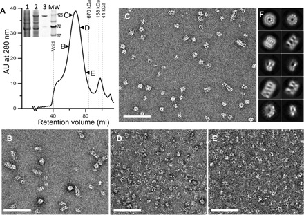
Fig. 1. Full-length wild-type MxB purified as oligomers.

(A) Purification of MBP-MxB from Expi293F cells by amylose affinity chromatography, followed by gel filtration through a Sephacryl S-500 HR column. The fractions indicated by arrowheads were visualized by negative stain EM. Inset, Coomassie-stained SDS–polyacrylamide gel electrophoresis (PAGE) gel of untransfected cells (1), transfected cells (2), and elution from amylose resin (3). Molecular weight (MW) markers are shown in kilodaltons (kDa). AU, absorbance units. (B to E) Representative negative stain micrographs of the respective fractions indicated in (A). (F) 2D class averages of the negatively stained MxB sample from fraction “C.” Scale bars, 200 nm.
