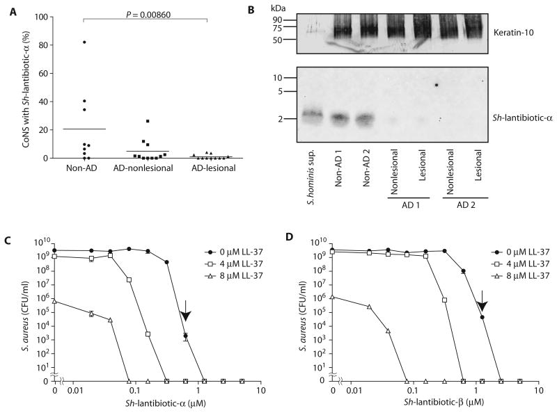
Fig. 6. Sh-lantibiotics are commonly found on healthy human skin and synergize with a host AMP.
(A) Frequency of detecting Sh-lantibiotic-α by colony PCR using gene-specific primers in CoNS isolates from human skin. Each point represents analysis of one individual. Bar, mean. P value was calculated by Wilcoxon-Mann-Whitney test. (B) Detection of Sh-lantibiotic-α peptide by Western blotting from extracts of skin swabs taken from two non-AD subjects who were colonized by bacteria having the Sh-lantibiotic-α gene and two AD subjects who were PCR-negative for the Sh-lantibiotic-α gene. S. hominis culture supernatant was loaded as a positive control. A total of 20 mg of protein was loaded in each lane. The uncropped image is shown in fig. S9. The membrane was restained with antibody against cytokeratin-10, a predominant protein in the stratum corneum, as a loading control. (C and D) Dose-response curves for the antimicrobial activity of Sh-lantibiotic-α (C) and Sh-lantibiotic-β (D) against S.aureus and their synergistic antimicrobial activity with human LL-37. Data represent means ± SEM of triplicate assays. Arrow shows minimal bactericidal concentration.

