
Fig. 6.
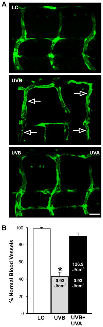
UVA-induced photo recovery in fli-1 zebrafish embryos. (A) Photomicrographs of the vasculature from a fli-1 larva that was not exposed to UV (Control, top), a fli-1 larva exposed to 0.93 J/cm2 UVB (middle), and a fli-1 larva that was exposed to 0.93 J/cm2 UVB followed by 126.9 J/cm2 UVA (bottom) during the mid-gastrulation stage of development. Open arrows point to abnormal ISBVs which projected in a straight trajectory along the dorsal–ventral axis. (B) Quantification in bar graph form indicates that the UVA exposure elicited photo recovery as 91 ± 4% of the ISBVs had normal morphology (LC = light control). Asterisk indicates significant difference (P < 0.05, Student’s T-Test) in the number of normal vessels between the UVB-exposed (n = 23) and the photo-recovered embryos (n = 22). Scale bar = 20 μm.