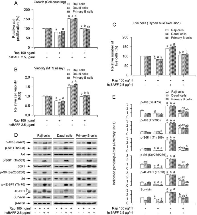
Fig. 1.

Inhibitory effect of administered rapamycin in Raji cells, Daudi cells and primary B lymphocytes on hsBAFF-stimulated phosphorylation of S6K1/4E-BP1 and Akt as well as cell proliferation and survival. Raji cells, Daudi cells and purified mouse splenic B lymphocytes were pretreated with/without rapamycin (Rap, 100 ng/ml) for 2 h, and then stimulated with/without 2.5 μg/ml hsBAFF for 12 h (for Western blotting) or 48 h (for cell proliferation and viability assay). (A) The cell proliferation was estimated by cell counting. (B) The cell viability was determined by the MTS assay. (C) The relative number of live cells was estimated by trypan blue exclusion assay. (D) Total cell lysates were subjected to Western blotting using indicated antibodies. The blots were probed for β-actin as a loading control. Similar results were observed in three independent experiments. (E) The blots for p-Akt (Ser473), p-Akt (Thr308), p-S6K1 (Thr389), p-S6 (Ser235/236), p-4E-BP1 (Thr70), and survivin were semi-quantified. Results are presented as mean ± SEM (n = 3–5). aP<0.05, difference with control group; bP<0.05, difference with 2.5 μg/ml hsBAFF group.
