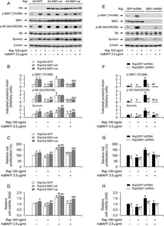
Fig. 4.

Effects of ectopic expression of wild-type or constitutively active S6K1 or down-regulation of S6K1 in Raji cells on rapamycin’s inhibition of hsBAFF-stimulated cell proliferation/viability. Raji cells, infected with Ad-S6K1-wt, Ad-S6K1-ca, or Ad-GFP (for control), were pretreated with/without rapamycin (Rap, 100 ng/ml) for 2 h, followed by stimulation with/without hsBAFF (2.5 μg/ml) for 12 h (for Western blotting) or 48 h (for cell proliferation and viability assay). (A and E) Total cell lysates were subjected to Western blotting using indicated antibodies. The blots were probed for β-actin as a loading control. Similar results were observed in at least three independent experiments. (B and F) The blots for p-S6K1 (Thr389), p-S6 (Ser235/236), and survivin were semi-quantified. (C and G) The cell proliferation was evaluated by cell counting. (D and H) The cell viability was determined by the MTS assay. Results are presented as mean ± SEM (n = 3–5). aP<0.05, difference with control group; bP<0.05, difference with 2.5 μg/ml hsBAFF group; cP<0.05, Ad-S6K1-wt group or Ad-S6K1-ca group vs Ad-GFP group; dP<0.05, S6K1 shRNA group vs GFP shRNA group.
