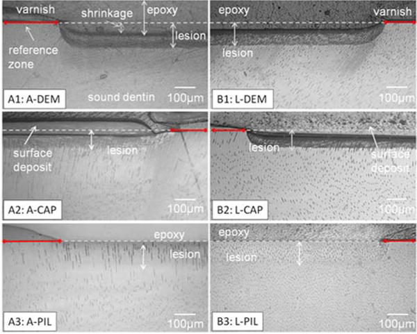
Fig.1.

Reflective light microscopy images of cross sections of artificial lesions before and after remineralization. Lactate took longer than acetate to create similar lesion depth. Collapse of severely demineralized substance was shown as a shrinkage when the specimens were embedded in dehydrating conditions (A1, B1). Artificial lesions created by both acetate and lactate showed full recovery of shrinkage after PILP remineralization for 14 days(A3,B3) whereas without PILP, there was less recovery (A2, B2).
