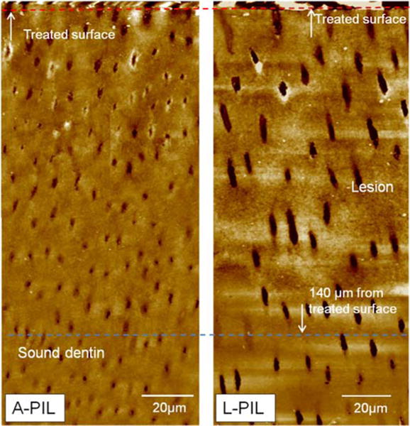
Fig.2.

AFM images of artificial dentin lesions created by acetate or lactate after PILP-remineralization for 14 days. Structures of both lesions were recovered, including some peritubular dentin. Dentin tubules in L-PIL look larger due to the tubule orientation in the cross section.
