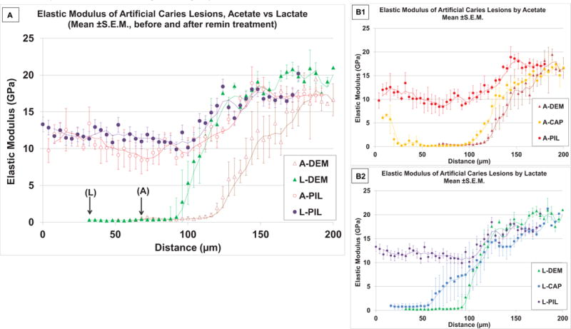
Fig.5.

Nanoindentation showing reduced elastic modulus (Er, n = 3) of artificial lesions from cross-sectioned specimens before and after PILP remineralization treatments. A) Comparison of Er along the lesion depth for two acids, (Acetate vs. Lactate), with arrows showing the actual measurement onset point due to shrinkage for each acid. Both acids had outer surface flat zone and inner sloped zone from the region of partial demineralization. After PILP remineralization, both lesions showed increases in Er in similar profiles. B1 and B2 show effects of PILP remineralization compared to the treatment without PASP in Er for each acid. Both acid lesions showed improved recovery at the outer flat zone when PASP was added to the remineralization solution. (Trendlines : moving averages).
