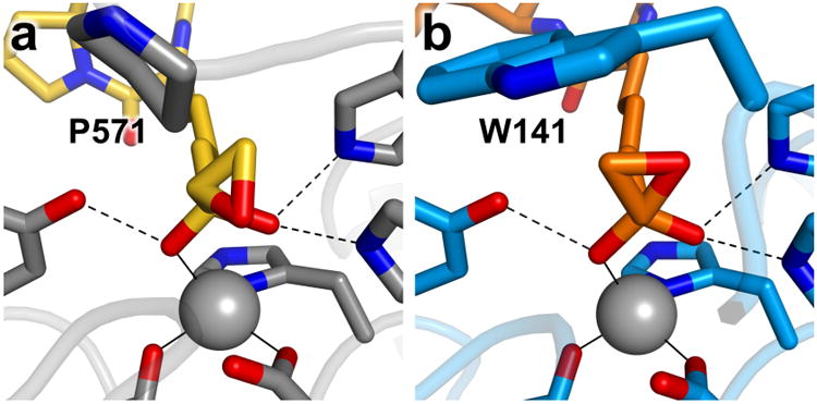
Figure 4.

Epoxyketone binding modes. (a) HC toxin (C = yellow, N = blue, O = red) bound to HDAC6 (C = gray, N = blue, O = red). (b) Trapoxin A (C = orange, N = blue, O = red) bound to HDAC8 (C = light blue, N = blue, O = red). Residues affecting epoxide conformations are labeled. Zinc ions are shown as gray spheres.
