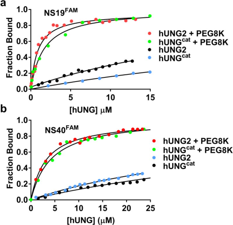
Figure 3.
Equilibrium binding of hUNG2 and hUNGcat to 19 and 40mer nonspecific DNAs in 150 mM [K+] in the presence and absence of 20% PEG8K (pH 7.5). (a) Changes in the fluorescence anisotropy of the DNA was used to measure binding of hUNG2 and hUNGcat to the 19mer duplex NS19FAM in the presence and absence of 20% PEG8K. (b) Binding of hUNG2 and hUNGcat to a 40mer duplex (NS40FAM) in the presence and absence of 20% PEG8K. The KD values in the presence of 150 mM [K+] and absence of PEG8K were very high (>35 µM). However, these were estimated by fixing the endpoint anisotropy to the value observed at low salt. The KD values for NS19FAM in the presence of PEG8K were 1.6 µM (hUNGcat) and 1.1 µM (hUNG2). The values for NS40FAM were 2.9 µM (hUNG2) and 3.6 µM (hUNGcat). All measurements were performed in triplicate.

