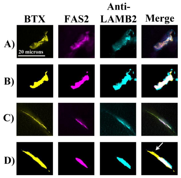
Figure 4. Typical staining patterns for an idiopathic scoliosis patient and the Escobar patient with the corresponding threshold images.
Skeletal muscle samples were triple stained for AChR (BTX; yellow), AChE (Fas2; magenta), and laminin β2 (cyan). Representative staining patterns of the three stains are shown. Threshold images for each stain were determined by the Image Pro software based on the number of pixels at each gray level (PLoS One, manuscript in revision). In the idiopathic scoliosis patient (A,B), the stains were highly colocalized as the formation of tightly apposed pre- and post-synaptic specializations at NMJs are hallmarks of neuromotor maturation. Compared to the idiopathic scoliosis patients, the Escobar patient (C,D) had more AChR present outside AChE (arrow) and less AChE present outside AChR.

