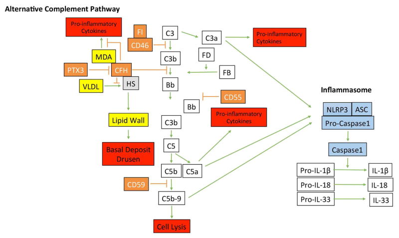
Figure 5.
Diagram of the alternative complement pathway and the inflammasome. Activation of the alternative complement pathway is regulated by factor H (CFH), CD46, CD55. PTX3, by binding CFH, controls the abundance of CFH. Besides its regulation of complement, CFH binds to malondialdehyde (MDA) to control MDA-mediated pro-inflammatory cytokine production, and heparan sulfates (HS) to compete with very low density lipoproteins (VLDL) that can become deposited in Bruch’s membrane prior to basal deposit or drusen formation. Complement activation can lead to anaphylatoxins generation of C3a and C5a, which can induce pro-inflammatory cytokine production, and C5b-9 complex formation, which is regulated by CD59. C3a, C5a, and C5b-9 complexes are implicated in activating the inflammasome, composed of NLRP3, ASP, and Pro-caspase1, which upon activation, is cleaved to convert pro-IL-1β, pro-IL-18, and pro-IL-33 into their active forms.

