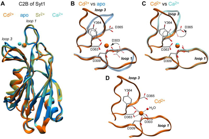
Figure 2.
Structural analysis of Cd2+-complexed C2B. (A) Backbone superposition of four C2B structures: Cd2+-complexed (this work, 5T0S, orange), apo penta-mutant (5CCJ,46 blue), Ca2+-complexed (1TJX,47 cyan), and Sr2+-complexed (1TJM,47 tan). (B, C) Pairwise comparison of loop regions of Cd2+-complexed C2B with those of the apo and Ca2+-complexed C2B highlights the differences between the conformation of loops and coordinating residues. The structural elements of apo and Ca2+-complexed C2B are shown with transparent representation. Only one Ca2+ ion and protein ligands of Cd2+ are shown for clarity in (C). (D) Coordination geometry of the Cd2+ site. Cd2+ has 7 oxygens ligands in its first coordination sphere, 6 contributed by C2B and 1 by a water molecule.

