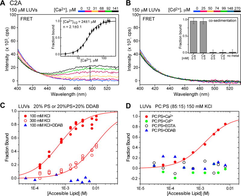
Figure 6.
Cd2+-complexed C2A domain does not associate with PtdSer-containing LUVs. (A) Ca2+-dependent fluorescence emission spectra showing an increase in the intensity of the dansyl band due to protein-membrane FRET. Inset: Ca2+-dependent C2A lipid-binding curve constructed using FRET intensity at 495 nm. (B) Cd2+-dependent fluorescence emission spectra demonstrating that no increase in dansyl emission intensity in the C2A–LUV system is observed upon addition of Cd2+. Inset: the results of vesicle sedimentation experiments that were conducted at 5 µM C2A and 1.5 mM total lipids. (C, D) C2A–lipid binding curves obtained using vesicle co-sedimentation experiments. The increase in ionic strength and neutralization of the negative charge by DDAB significantly weakens the interaction between Ca2+-complexed C2A and membranes (C). No binding of Cd2+-complexed C2A to PtdSer-containing vesicles is observed (D). In both (C) and (D), Ca2+ and Cd2+ are added to a concentration of 1 mM.

