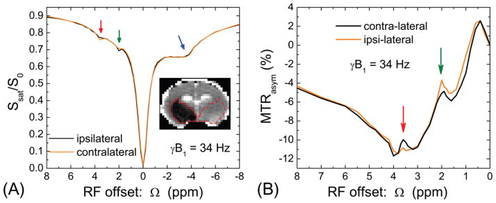
Fig. 7. In vivo Z-spectra and MTRasym-spectra from rat brain with MCAO.
Z-spectra with a saturation power of 34 Hz were obtained from rat brains with ischemic region (n = 4). The ipsilateral (solid contour) and contralateral (dashed contour) ROIs were selected based on the ADC map (Inset). (A) Z-spectra obtained from two ROIs are similar for most of the frequency range, except small changes at the amide and guanidyl frequencies. (B) The MTRasym-spectra showed that the ipsilateral ischemic region has reduced amide-CEST and enhanced guanidyl-CEST signals. Note that other signals in the Z-spectra are insensitive to ischemia, such as from aliphatic protons or the MTC effect.

