
Figure 3.
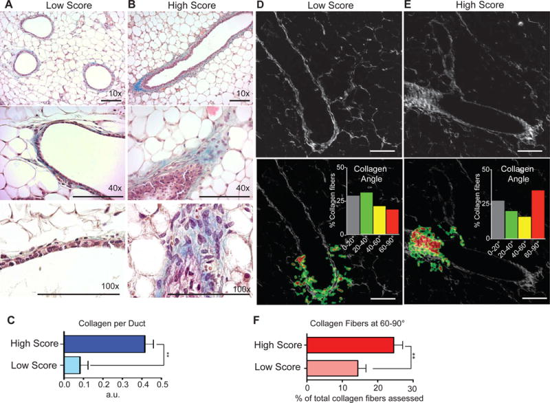
Exposure of normal mammary gland to high invasion score PSF promotes hyperplasia, collagen deposition, and formation of tumor associated collagen signature 3 (TACS3). A) H&E and Masson’s Trichome stain of mammary gland exposed to 1% low score PSF. B) H&E and Masson’s Trichome stain of mammary gland exposed to 1% high score PSF. C) Quantification of collagen staining (p = 0.0032; n = 3 low score, n = 4 high score, Unpaired t-test with Welch’s correction). D) Second harmonic generation imaging and CurveAlign software analysis of mammary ducts exposed to low or high score PSF. (scale bar = 100μm). E) The same was done for high score PSF treated mammary glands identifying the majority of collagen fibers interacting with the ductal border at 60–90°, the empirical definition for TACS3 conformation (scale bar = 100μm). F) Quantification of TACS3 in mammary glands exposed to low and high score PSF (p = 0.0163; n = 4 low score, n = 7 high score, Unpaired t-test with Welch’s correction).