
Figure 1.
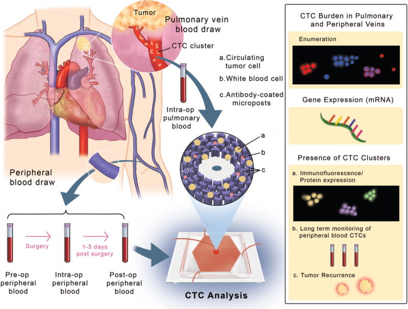
Study schematic representing the anatomy of the lung displaying the pulmonary vein (PV) and peripheral veins (Pe). Blood was drawn from the veins at different time-points around tumor resection and processed through the OncoBean Chip, followed by analysis by enumeration and genomic profiling (inset on right).