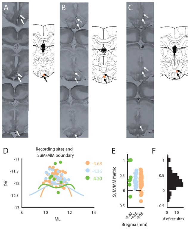
Figure 1.

Anatomical reconstruction and distribution of recording sites within the mammillary (MM) area. (A) Raw histological sections (left) and reconstructed recording site (right) in the MM. (B) Raw histological sections (left) and reconstructed recording site (right) in the SuM/MM border. (C) Raw histological sections (left) and reconstructed recording site (right) in the SuM. (D) Anatomical boundaries between the SuM and MM with putative recording sites across three anterior-posterior positions according to the brain atlas. (E) Distribution of recording sites as per our SuM/MM metric. (F) Total distribution of recording sites across our SuM/MM metric. DV, Dorso-ventral; ML, Medio-lateral; SuM, supramammillary nucleus; MM, mammillary bodies.
