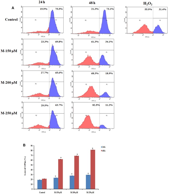
FIGURE 4.
Quantification of loss of mitochondrial membrane potential by Rhodamine 123 staining. SW480 cells were treated with various concentrations of morin (150, 200, and 250 μM) for 24 and 48 h and H2O2 (200 μM) for 2 h. After incubation, cells were stained with Rhodamine 123 analyzed using flow cytometer. (A) Representative results. (B) Data analyzing fluorescence intensity from triplicate measurements. M (150, 200, and 250 μM): Morin (150, 200, and 250 μM). Significance levels between different groups were determined by using one way ANOVA, followed by Duncan’s multiple range test. ∗p ≤ 0.05 versus control.

