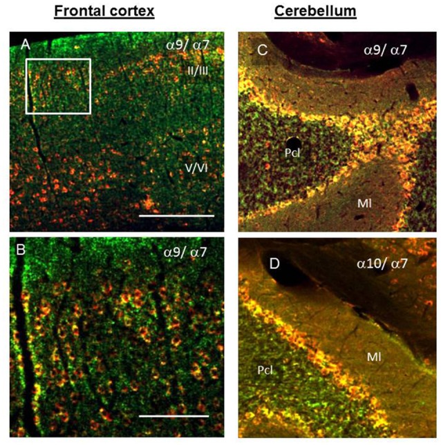
Figure 7.

The presence of α7 (green), α9 and α10 (red) nAChR subunits in the II/III and V/VI layers of SC (A,B, insert); cortical layers specification according to Ahissar and Staiger, 2010) and cerebellum (C,D). Pcl, Purkinje and granule cell layers; Ml, molecular layer. Yellow—merge of the red and green staining. In (A,C,D) bar is 200 μm, in (B) 100 μm.
