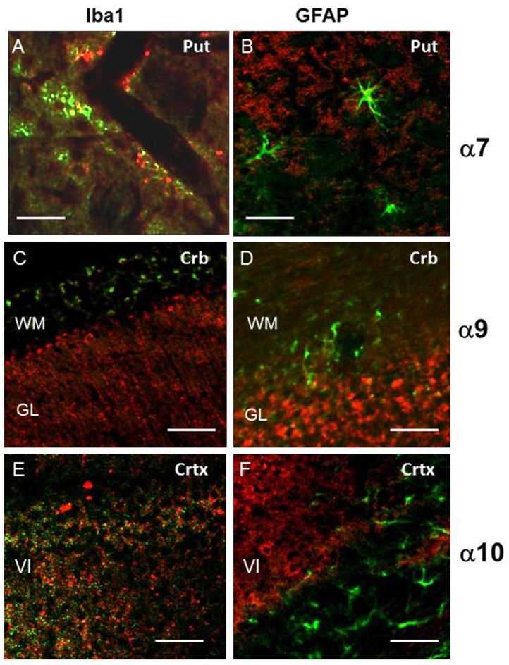
Figure 8.
The double staining for α7, α9 or α10 nAChR subunits (red) and markers of microglia (Iba1, green, A,C,E) or astrocytes glial fibrillary acidic protein (GFAP, green, B,D,F) in the cerebellum (Crb), cortex (Crtx) or putramen (Put). Abbreviations: WM, white matter; GL, granular layer of cerebellum; V, internal pyramidal layer; VI, multiform layer of cortex. In (A) bar is 20 μm, in (B–F) 50 μm.

