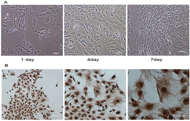
Figure 1. Immunohistochemistry and morphology identify of mandibular condylar chondrocytes.

Normal condylar chondrocytes special expressed COL2. (A) The morphology of normal chondrocytes cultured for 1, 4and 7 days was observed under a microscope. (B) The immunohistochemical staining shown that COL2 was positive in the chondrocytes.
