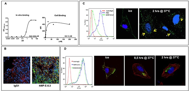
Figure 1.

hMP-E-8. 3 binds endosialin in vitro/in vivo and is internalized into SJSA-1 cells (A) Binding of hMP-E-8.3 to human recombinant endosialin's ECD by ELISA (left) or FACS analysis on living osteosarcoma SJSA-1 cells (right). (B) Binding of hMP-E-8.3 to endosialin in vivo as evaluated by immunofluorescence analysis of SJSA-1 tumor xenografts. Animals received a single injection of human IgG (as a control), or hMP-E-8.3, both at the dose of 10 mg/Kg. Twenty-four later, animals were sacrificed, the tumors frozen and tumor sections stained with AlexaFluor-488 conjugated anti-human IgG (green), a commercial antibody against human endosialin followed by AlexaFluor-546 conjugated anti-rabbit antibody staining (red) or Draq5 (blue). (C) Flow cytometry analysis (left panel) of endosialin surface expression in SJSA-1 cells cultured in the presence of hMP-E-8.3 (10 μg/ml) at 37°C for 2 hrs. The antibody is efficiently internalized as revealed by a marked reduction of endosialin expression (85% in 2 hrs). Fluorescence immunocytochemistry (right panels) of hMP-E-8.3 internalization in SJSA-1 cells. Cells were incubated with hMP-E-8.3 on ice for 20 min, then shifted at 37°C for 2 hrs. After harvesting, cells were stained with AlexaFluor-488 conjugated anti-human IgG (green). Draq5 (blue) was used to visualize nuclei. Yellow arrows indicate intracellular staining. Images were acquired with LSM-510 laser scanning confocal microscopy. (D) Flow cytometry analysis (left panel) of endosialin surface expression in SJSA-1 cells as evaluated by naked hMP-E-8.3 antibody or ENDOS/ADC. Confocal imaging (right panels) of ENDOS/ADC internalization in SJSA-1 cells. Cells were incubated with ENDOS/ADC (10 μg/ml) on ice for 20 min, then shifted at 37°C for the indicated times. After harvesting, cells were stained with AlexaFluor-488 conjugated anti-human IgG (green), Rhodamine-labeled phalloidin was used to visualize actin cytoskeleton (red), Draq 5 (blue) for nuclei staining.
