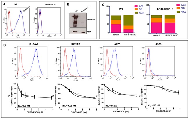
Figure 2. ENDOS/ADC in vitro cytotoxic activity correlates with endosialin expression.

Endosialin −/− SJSA-1 cells were generated by CRISPR-Cas9 system of genome editing. Lack of endosialin expression was documented by (A) FACS analysis and (B) Western blotting. (C) Cell cycle analysis was performed after exposure of wild-type or endosialin −/− SJSA-1 cells to 0.4 μg/ml of ENDOS/ADC. (D) Levels of endosialin surface expression determined by cytometry analysis using hMP-E-8.3 antibody for cells staining (upper panel). ENDOS/ADC cell killing activity (lower panel) after 120hrs of cells exposure to increasing doses of ENDOS/ADC. The following human cancer cell lines and tissue of origin were used: SJSA-1, osteosarcoma; SKNAS, neuroblastoma; A673 Ewing's sarcoma; A375 melanoma. The values are expressed as mean ± SD of three experiments. IC50 and best-fit curve were calculated using GraphPad Prism 5 software.
