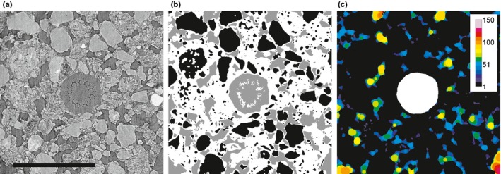
Figure 2.

(a) Cross‐section of barley root (no hairs) growing in soil. Internal root structures and the surrounding soil structure could be clearly visualised. Bar, 1 mm. (b) Soil classification using trainable WEKA segmentation. Black, solid phase; white, mixed phase; grey, air‐filled pore space. Note that the root was segmented independently. (c) Pore size classification around the root. Segmented root is shown in white. Colours indicate local pore diameter in micrometres.
