
Figure 3.
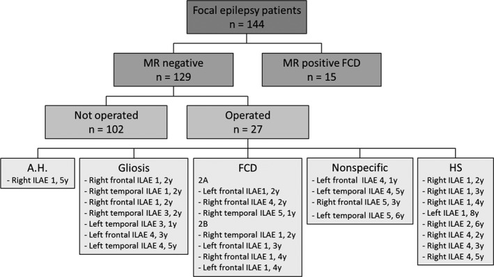
Study tree. This figure gives an overview of the patients included in this study, comprising 144 focal epilepsy patients, including 15 epilepsy patients with a preoperatively diagnosed and postoperatively histopathologically proven focal cortical dysplasia (FCD) and 129 epilepsy patients who had normal magnetic resonance imaging (MRI). Twenty‐seven of these patients had epilepsy surgery, of whom seven had a histopathologically proven FCD, eight had hippocampal sclerosis (HS), seven had gliosis, four were nonspecific and one had amygdala hamartoma (A.H.). The postoperative outcome is indicated based on the International League Against Epilepsy (ILAE) classification 1–6; a good outcome was defined as ILAE classification = 1–2, poor outcome as ILAE classification = 3–6. In total, 14 had a good (ILAE classification = 1–2), 13 a poor (ILAE classification = 3–6) postoperative outcome at 1–7 years (average follow‐up = 3.3 years). The duration of postoperative follow‐up for each patient is indicated following the ILAE classification.