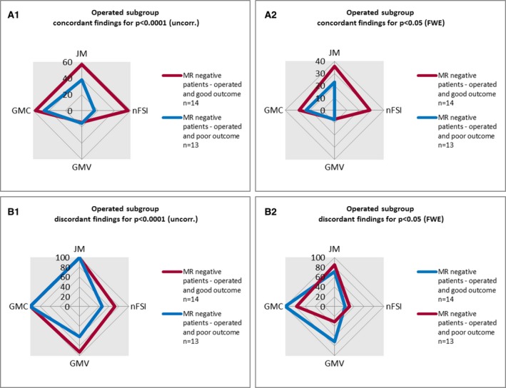
Figure 5.

Graphical overview of voxel‐based morphometry (VBM) findings in the subgroup of operated magnetic resonance (MRI)‐negative patients. This figure summarizes the VBM findings in the MRI‐negative subgroups with good (International League Against Epilepsy [ILAE] classification = 1–2) and poor (ILAE classification = 3–5) postsurgical outcome for each map (gray matter concentration [GMC], gray matter volume [GMV], junction map [JM], nFSI = normalized T2–fluid‐attenuated inversion recovery [nFSI]) at two different thresholds: (A1, B1) p < 0.0001 (uncorrected [uncorr.]); (A2, B2) p < 0.05 (family‐wise error rate [FWE]).
