
Figure 2.
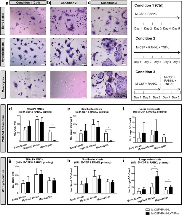
Effect of TNF‐α on osteoclastogenesis by different osteoclast precursors on plastic. (a–c) Early blasts (1st row), myeloid blasts (2nd row), and monocytes (3rd row) were cultured under three different culture conditions: (a) condition 1 (control), with 30 ng/ml M‐CSF and 20 ng/ml RANKL for 5 days; (b) condition 2, with 30 ng/ml M‐CSF, 20 ng/ml RANKL and 10 ng/ml TNF‐α for 5 days; (c) condition 3, first cultured with 30 ng/ml M‐CSF and 20 ng/ml RANKL for 3 days, then cells were further cultured with 30 ng/ml M‐CSF, 20 ng/ml RANKL and 10 ng/ml TNF‐α for 2 days. Cells were stained for TRAcP activity and nuclei were counterstained by DAPI. Osteoclasts were recognized as TRAcP+ multinucleated cells (purple) and nuclei were visualized as blue. Note the absence of any multinucleated cells in monocyte cultures in condition 2. Scale bar = 100 μm. (d–f). Total number of TRAcP+ multinucleated cells (>2 nuclei), number of small osteoclasts (3–10 nuclei), and number of large osteoclasts (>10 nuclei) generated by the three subsets. White column: cells cultured with 30 ng/ml M‐CSF and 20 ng/ml RANKL for 5 days (condition 1). Black column: cells cultured with 30 ng/ml M‐CSF, 20 ng/ml RANKL, and 10 ng/ml TNF‐α for 5 days (condition 2). (g–i). Total number of TRAcP+ multinucleated cells (>2 nuclei), number of small osteoclasts (3–10 nuclei), and number of large osteoclasts (>10 nuclei) generated by the three subsets with M‐CSF and RANKL priming. White column: cells cultured with 30 ng/ml M‐CSF and 20 ng/ml RANKL for 5 days (condition 1). Black column: cells first cultured with 30 ng/ml M‐CSF and 20 ng/ml RANKL for 3 days, then 10 ng/ml TNF‐α was added and cells were further cultured with M‐CSF, RANKL, and TNF‐α for 2 more days (condition 3). (n = 6, *p < 0.05, **p < 0.01, ***p < 0.001)