
Figure 5.
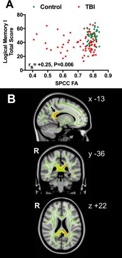
Relationship between recall memory and splenium fractional anisotropy (FA) in patients after traumatic brain injury (TBI) and controls. (A) Relationship between mean splenium of corpus callosum (SPCC) FA as independent variable and Logical Memory I (LMI) total score (immediate recall) as dependent variable, in patients after moderate–severe TBI (n = 82, red circles) and controls of similar age and sex (n = 40, green circles), without adjustment for any covariates. rS represents Spearman correlation coefficient for combined groups. This represents a separate cross‐sectional cohort of subjects to the longitudinal cohort. (B) Group average statistical maps showing voxels (in red) in SPCC (arrows) and inferior longitudinal fasciculus (arrowhead) white matter (WM) tracts whose FA is positively correlated with LMI total score (immediate recall) in TBI patients (n = 82). Results are overlaid on combined group FA skeleton (green, threshold > 0.2), using threshold‐free cluster enhancement clusterwise threshold p < 0.05, correcting for age and sex. Results are displayed on standard Montreal Neurological Institute 125 (MNI125) 1mm T1 magnetic resonance imaging brain scan. SPCC WM tract defined using the Johns Hopkins University WM tractography atlas with a cutoff threshold of 20% is indicated in yellow. Mean FA values in the SPCC WM tract region of interest in A were calculated from the average of the voxels indicated by the overlap of the green skeleton and yellow mask. Coordinates are given in MNI standard space. R = right.