
Fig 2.
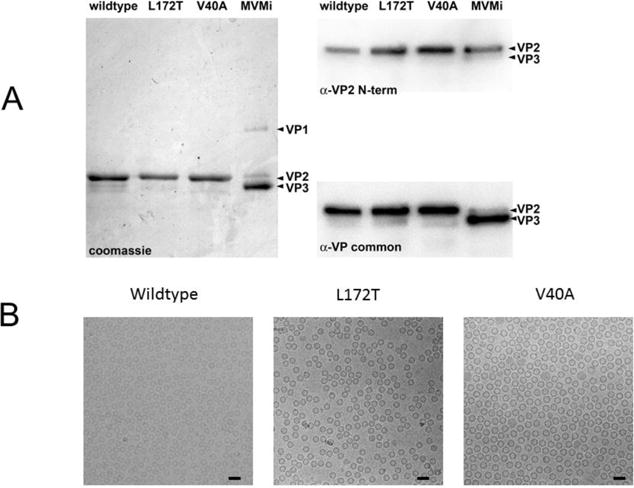
(A) The left panel shows a Coomassie Brilliant Blue-stained SDS-PAGE of total protein present in samples of purified MVMi wildtype, L172T and V40A VP2-only VLPs, run in parallel with authentic MVMi virions. The latter show the positions of VP1 and VP2, and of VP3, the proteolytic cleavage product found in DNA-containing virions, which has lost ~25 amino acids from its N-terminus. The top right panel shows a western blot of the VP2/VP3 region of a parallel SDS-PAGE probed with α-VP2 N-term, a rabbit polyclonal antibody raised against a 25mer peptide with the sequence NH2 - M S D G T S Q P D G G N A V H S A A R V E R A A - COOH, corresponding to residues 1 to 24 of the VP2 sequence. The bottom right panel shows the same western blot probed with α-VP common, a rabbit polyclonal antibody raised against an 18mer oligopeptide with the sequence NH2 - Q G S R H G A T Q M E V N W V S K - COOH, located in the MVMi VP sequence from amino acid residues 453 to 469 of VP1 and 311 to 327 of VP2. (B) Cryo-EM micrographs of vitrified MVM show (left to right) assembled wildtype, L172T, and V40A VLPs (50 nm scalebar).