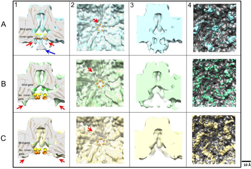
Fig 4.

In panel 1, maps of wildtype (A-1), L172T (B-1), and V40A (C-1) VLPs were surface rendered and a central cross-section view of the five-fold channel is shown with the alpha carbon trace from the crystal structure (PDB ID 1Z14) fitted into the cryo-EM densities. The shape of the pore in L172T cuts back sharply from the enhanced mid-gate constriction, leaving the fitted L172 residue (green spheres) out of density compared to the wildtype and V40A VLPs. Below the mid-gate, the base of the pore in V40A also opens, so that the fitted residues G39 (orange spheres) and V40 (yellow spheres) are out of density, and the inner constriction of the channel is lost. In the wildtype map, the five-fold density funnel (blue arrow in panel A-1) has magnitude as strong as the capsid density, and remains strong even at higher contour levels at which some capsid densities disappear. In panel 2, the five-fold vertex is viewed along the five-fold axis from the interior of the capsid. Claw-like densities surrounding the base of the five-fold pore (red arrows) are rendered at a density contour of 1 sigma. Panel 3 compares maps at lower contour levels (sigma = 0.6), where more flexible densities can be seen. Here, the claw-like features merge with the funnel density in the wildtype map (blue). Although claw-like densities were seen for both L172T (green) and V40A (yellow), compared to wildtype they are in a different location, laterally displaced away from the five-fold axis. Panel 4 shows the difference densities between the MVM crystal structure (PDB ID: 1Z14) rendered as a density map and the cryo-EM maps of wildtype, L172T and V40A VLPs. The difference densities were segmented and the density around the five-fold were quantified. The volume of the wildtype, L172T and V40A claws were 697, 1287 and 1762 respectively (not shown).
