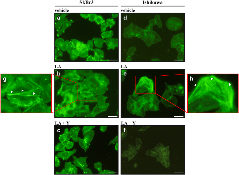
Figure 6.
Lauric acid promotes the formation of stress fibers. SkBr3 and Ishikawa cells were treated for 4 h with vehicle (−) (a, d) or 100 μM LA alone (b, e) or in combination with 10 μM ROCK inhibitor Y-27632 (Y) (c, f) and subjected to phalloidin staining to visualize F-actin. (g, h) Enlarged details of stress fibers shown in b and e, respectively. White arrows indicate stress fibers. Images shown are representative of 30 random fields obtained in three independent experiments. Scale bar: 12.5 μm.

