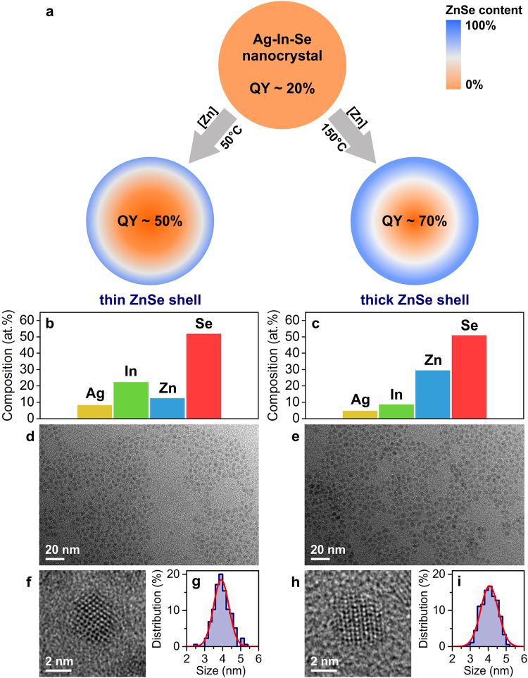
Figure 2.
Model system: graded Ag-In-Se/ZnSe core/shell nanocrystals. (a) Schematic illustration of the ZnSe shell growth process. Two investigated samples with distinct ZnSe atomic profiles are prepared at different shell growth temperatures. QY denoted luminescence quantum yield. (b,c) Energy dispersive X-ray spectra quantifications, (d,e) representative TEM images, (f,h) high-resolution TEM images, and (g,i) size distributions of investigated Ag-In-Se/ZnSe core/shell nanocrystals. Data for thin ZnSe shell sample are shown in the left panel and for thick ZnSe shell sample in the right panel.

