
Fig. 1.
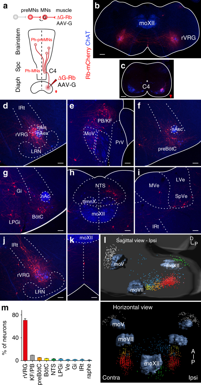
Distribution of Ph-preMNs in P9 mice following unilateral viral injections of the diaphragm. a Monosynaptic tracing scheme for Ph-preMNs using a G-deficient Rb virus (ΔG-Rb) and a G-coding adeno-associated (AAV-G) viral cocktail. b, c Brainstem and cervical spinal transverse sections showing, respectively, bilaterally distributed trace+ Ph-preMNs of the rVRG (b) and ipsilaterally located seeding trace+ Ph-MNs. d–k Representative images of transverse sections of the brainstem showing (in decreasing abundance order) trace+ Ph-preMNs in the rVRG (d), in the PB/KF (e), in the preBötC (f), in the BötC, Gi and LPGi (g), in the NTS (h), in Ve nuclei (i), in the lRt (j), and in the raphe (k). l 3D reconstructions of the brainstem showing the spatial distribution of Ph-preMNs (n = 4 cumulated counts) in sagittal (top) and horizontal (bottom) views (color code of locations is given in m). m Summary histogram of the distribution Ph-preMNs (percent of total trace+ cells). dmnX, dorsal motor nucleus of the vagus, LRN, lateral reticular nucleus; LVe, lateral vestibular nucleus; MoV, trigeminal motor nucleus; MoXII, hypoglossal motor nucleus; MVe, medial vestibular nucleus; nAc, compact nucleus ambiguus; nAex, external formation of nucleus ambiguus; nAls loose formation of nucleus ambiguus; nAsc, semi-compact nucleus ambiguus; PrV, principal sensory trigeminal nucleus; Spc, spinal cord; SpVe, spinal vestibular nucleus. Scale bars b, c 200 µm; d–k 100 µm