
Fig. 2.
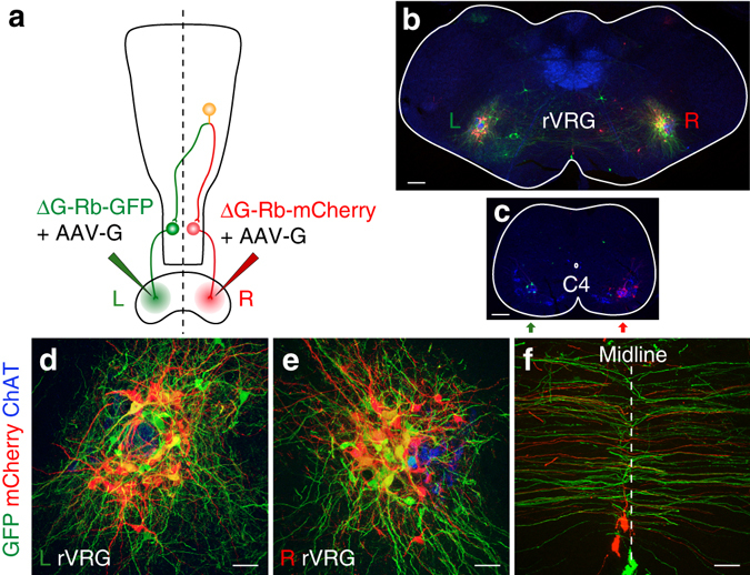
Individual Ph-preMNs in the rVRG project bilaterally on Ph-MNs. a Tracing scheme based on injections of a green virus (Rb-GFP) in the left diaphragm (L green lettering) and of a red virus (Rb-mCherry) in the right diaphragm (R red lettering). b, c Transverse sections at the level of the rVRG (b) and at the C4 level (c). Note the presence of double labeled (GFP+/mCherry+, yellow) rVRG neurons on the left and right side (b) while seeding Ph-MNs (c) on each side express exclusively either GFP (green) or mCherry (red). d–f Close-up view of the left (d) and right (e) rVRG showing exclusive green or red cells as well as double labeled cells (yellow). f Close-up view of labeled commissural axons over the midline (dotted line). Scale bars: b, c 200 µm; d–f 50 µm