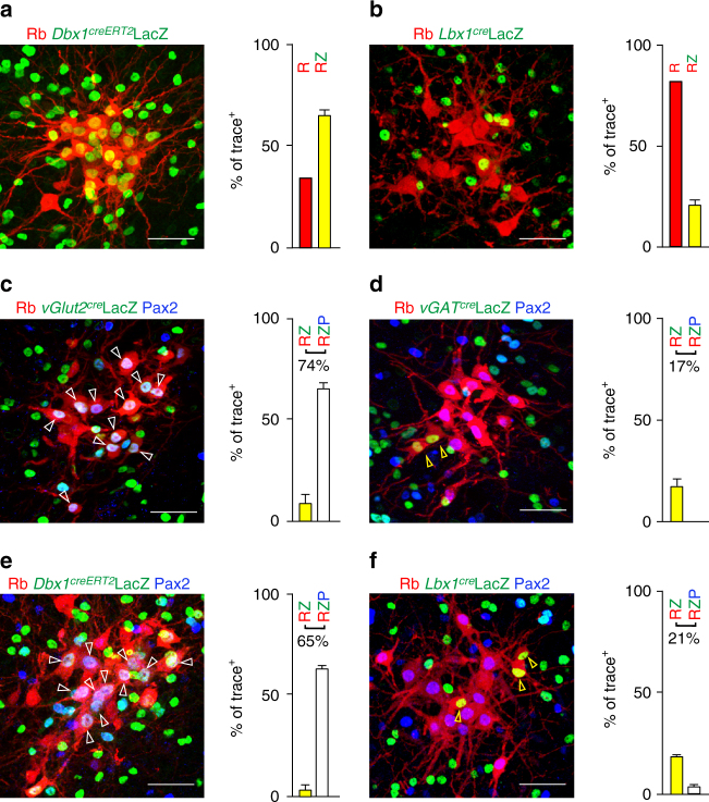
Fig. 3.
Identity of the Ph-preMNs of the rostral ventral respiratory group. a, b Transverse brainstem section showing trace+ rVRG neurons labeled by Rb-mCherry (red, R) and counterstained for nuclear expression of LacZ (green, Z) and summary histograms featuring the percentage of trace+ rVRG neurons expressing LacZ (yellow bars, RZ). The rVRG is comprised of neurons with a history of expression of Dbx1 (a) or Lbx1 (b). c–f Same as above with additional immunostaining for Pax2 (blue, P) and summary histograms showing the percentage of trace+ rVRG neurons expressing LacZ alone (yellow bars, RZ, yellow arrowhead) or co-expressed with Pax2 (white bars, RZP, white arrowhead) when LacZ is expressed from the vGlut2 locus (c); from the vGAT locus (d); from the Dbx1 locus (e) and from the Lbx1 locus (f). Note the comparable proportion of triple positive cells in c, e panels, of double positive cells in d, f panels and the virtual absence of triple positive cells in d, f panels. Scale bars: 50 µm

