
Fig. 4.
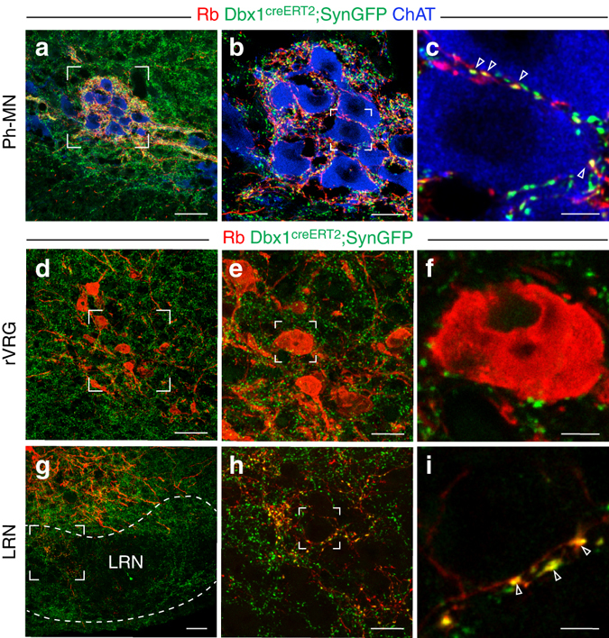
Synaptic targets of V0 rVRG neurons. a V0 rVRG synaptic terminals double labeled (yellow) by rabies-mCherry (Rb, red) and SynGFP (green) are massively present on Ph-MNs (trace−, ChAT+, blue) contralateral to the diaphragm viral injection side. b Zoom on the square inset in a. c Single optical section of the inset in b showing individual synapses (arrowheads). d–f rVRG (trace+, red) neurons are devoid of double labeled terminals. e Zoom of the inset in d. f Single optical section of the inset in e. g V0 trace+ rVRG neurons project to the lateral reticular nucleus (LRN). h Zoom of the inset in g. i Single optical section of the inset in h showing individual V0 rVRG synaptic terminals (arrowheads) presumably abutting the soma of a LRN neuron. Scale bars: a, d, g 50 µm; b, e, h 20 µm; c, f, i 5 µm