Fig. 6.
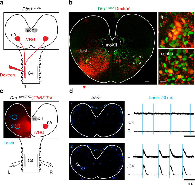
Neurons of the rVRG transmit bilaterally their excitation to Ph-MNs at E15.5. a Retrograde tracing scheme in a Dbx1 LacZ/+ brainstem spinal cord at E15.5 using Rhodamine dextran dye unilaterally injected in at C4 level. b Left, transverse slice showing the tracer pattern (red) and LacZ counterstain (green) in the ipsi- and contralateral rVRG (insets) to the tracer injection side. Right, zoom on the insets showing double labeled (yellow) rVRG cells. c Schematic showing the Dbx1 creERT2 ; ChR2-Tdt preparation and unilateral illumination targets (blue empty circles) achieved by computer generated holography outside (1) and on (2) the rVRG while recording activities of left and right C4 motor roots. d Top photostimulation away from the rVRG (blue circle 1 in c) fails both to trigger detectable ∆F/F fluorescence changes and evoke C4 activity responses (right set of traces). Bottom, the light spot positioned on the rVRG (blue circle 2 in c) evokes ∆F/F fluorescence changes restricted to the targeted rVRG (arrowhead) and synchronous left and right C4 bursts (right set of traces). Note that the photostimulation of the rVRG on one side fails to activate the contralateral rVRG. Scale bars: b left, 100 µm; b right, 20 µm

