Figure 4.
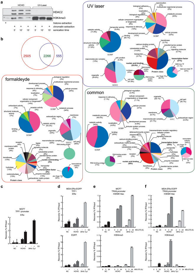
UV laser induces DNA-protein binding. (a) Western blotting analysis on chromatin derived from formaldehyde- and UV laser-treated cells. Histone extract was used as internal control. (b) Venn diagram showing number of proteins identified by MS/MS analysis within chromatin complexes derived from formaldehyde (red)- and laser (blue)-treated cells. Rectangles contain GO term analyses of each protein (formaldehyde = red, UV laser = blue and common proteins = violet). (c and d) ChIP assays performed in indicated cells treated with E2 and crosslinked with formaldehyde and UV laser showing TFF1 promoter region occupancy by (c and d, top) ERα and (d, bottom) GFP. (e,f) ChIP assays performed in indicated cells treated with MS-275 at indicated times and crosslinked with formaldehyde or UV laser showing TRAIL promoter region occupancy by H3K9K14ac (e,f, top) and H3K4me3 (e and f, bottom). All ChIP data obtained on immunoprecipitated fractions were normalized to input chromatin (IP/Input). Curves show the mean of at least two independent experiments with error bars indicating standard deviation.

