Figure 1.
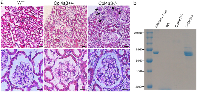
Analysis of proteinuria and kidney histology. (a) Representative images of H&E staining of WT, Col4a3+/−, and Col4a3−/− kidney sections. Glomeruli in wild-type (WT) and Col4a3+/− kidneys have a normal appearance. The right upper panel shows several sclerosed glomeruli (arrows) along the surface of a cortical lesion and interstitial injury in a Col4a3−/− kidney. Scale bars, 50 μm in upper panels; 10 μm in lower panels. (b) Coomassie blue staining reveals a strong albumin band in spot urines collected from Col4a3−/− mice but not from the WT and Col4a3+/− mice.

