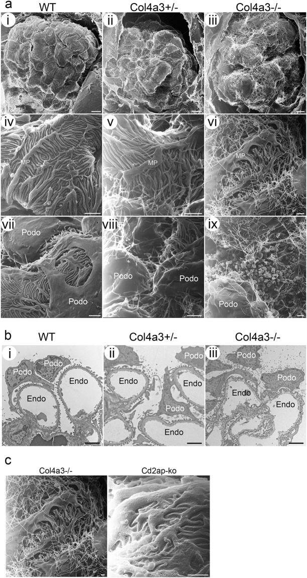
Figure 2.
Glomerular and podocyte morphology in wild-type and Col4a3 mutant mice. (a) HIM images of glomeruli in wild-type (WT) (i,iv,vii), Col4a3+/− (ii,v,viii), and Col4a3−/− mice (iii,vi,ix). Low magnification image of WT kidney (i) shows the glomeruli formed of capillary loops covered with podocytes while images of Col4a3+/− (ii) and Col4a3−/− kidneys (iii) show a less regular and less smooth surface of podocytes. High magnification images of WT kidneys (iv,vii) show multiple thick primary or major foot processes projecting from the podocyte cell body and covering the capillary loops. The primary processes frequently cross over each other, and numerous uniformly shaped underlying foot processes arise from the primary processes forming an interdigitated network. There are rounded and filamentous microprojections on the free surface of the podocyte cell body and primary processes (vii). The primary processes are flattened, broader and less organized, and the foot processes more randomly branched in Col4a3−/− kidneys (vi) compared to WT. An increased number of longer microprojections, arising from primary processes and foot processes, was detectable in Col4a3+/− (v,viii) and Col4a3−/− kidneys (vi,ix). Numerous “blebs” projecting out of the epithelial surface between podocyte cell bodies could be observed (ix). Scale bars, 5 μm in upper panels; 1 μm in middle panels and lower panels. (b) Representative TEM images of WT (i), Col4a3+/− (ii), and Col4a3−/− glomeruli (iii). WT glomeruli (i) show normal GBM thickness and regularly lined-up foot processes. Col4a3−/− glomeruli (iii) show irregularly thickened and lamellated GBM. Some podocytes have widened foot processes and SDs disappear between interdigitating foot processes. Increased microvilli formations are detectable. Scale bars, 2 μm. MP, major process; Podo, podocyte; Endo, endocapillary space. (c) Microprojections in Col4a3 and Cd2ap knockout (ko) mice. HIM images of glomeruli in Col4a3−/− (left panel, copied from Fig. 2a-vi, for comparison) and Cd2ap-ko mice (right panel). While an increased number of longer microprojections arise from primary processes and foot processes in Col4a3−/− kidneys, they are less obvious in Cd2ap-ko kidneys. Scale bars, 500 nm.

