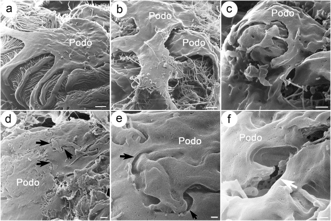
Figure 5.
Podocyte ultrastructure at late stage in Col4a3 mutant mice compared with wild-type mice. Shown are HIM images of podocytes at late stage in wild-type (WT) (a) and Col4a3−/− mice (b–f). (a) WT podocytes show intact podocyte structure with major processes and foot processes forming interdigitated structures. Late stage Col4a3−/− podocytes show broadly effaced podocyte processes that form large flattened sheets covering capillary walls (b,c). In some regions, podocytes appear connected by junctional structures (arrows) (d,e). In other regions, the presence of a few breaks/holes and gaps between podocytes is detected (white arrow) (f). A mesh structure appearing as degraded endothelial fenestrae is visible in the gap. Scale bars, (a,b,d) 1 μm; (c) 500 nm; (e,f) 200 nm. Podo, podocyte.

