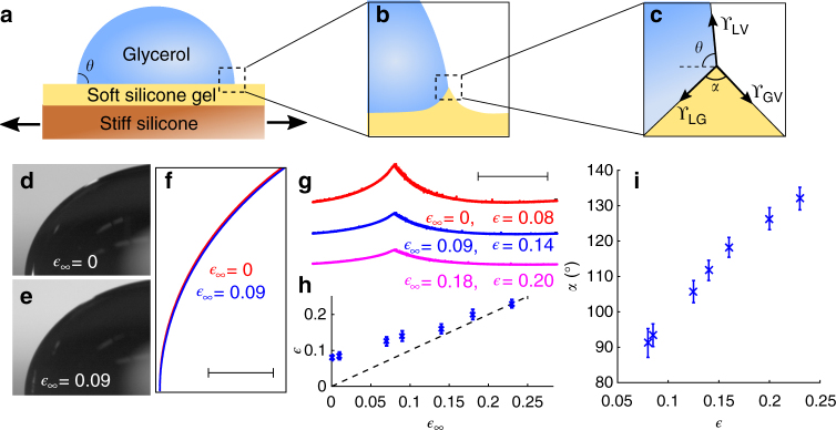
Fig. 2.
Macroscopic contact angle and microscopic wetting profiles. a Schematic of the strain-dependent wetting experiments, using a biaxial stretcher as described in ref. 50. b Detail of the contact line geometry at intermediate scales. c Detail of the contact line at microscopic scales, much less than ϒ/E. At this scale, the geometry of the contact line is given by a vector balance of the surface stresses as shown. d, e Macroscopic wetting profiles of large glycerol droplets sitting on unstretched and stretched ( = 0.09) silicone gels. f Superimposed boundaries for the drops on the stretched (blue) and unstretched (red) substrates show no difference in the macroscopic contact line geometry (scale bar: 400 μm). g Microscopic wetting profiles for a single droplet on unstretched (red), 9% stretched (blue), and 18% stretched (pink) silicone gel substrates, respectively (scale bar: 20 μm). h Local strain near the contact point, , plotted against the applied strain, . Dashed line has a slope of 1. i The opening angle of the wetting ridge, α, increases with the local strain, . In h, i, the error bars are SD of the population

