Figure 6.
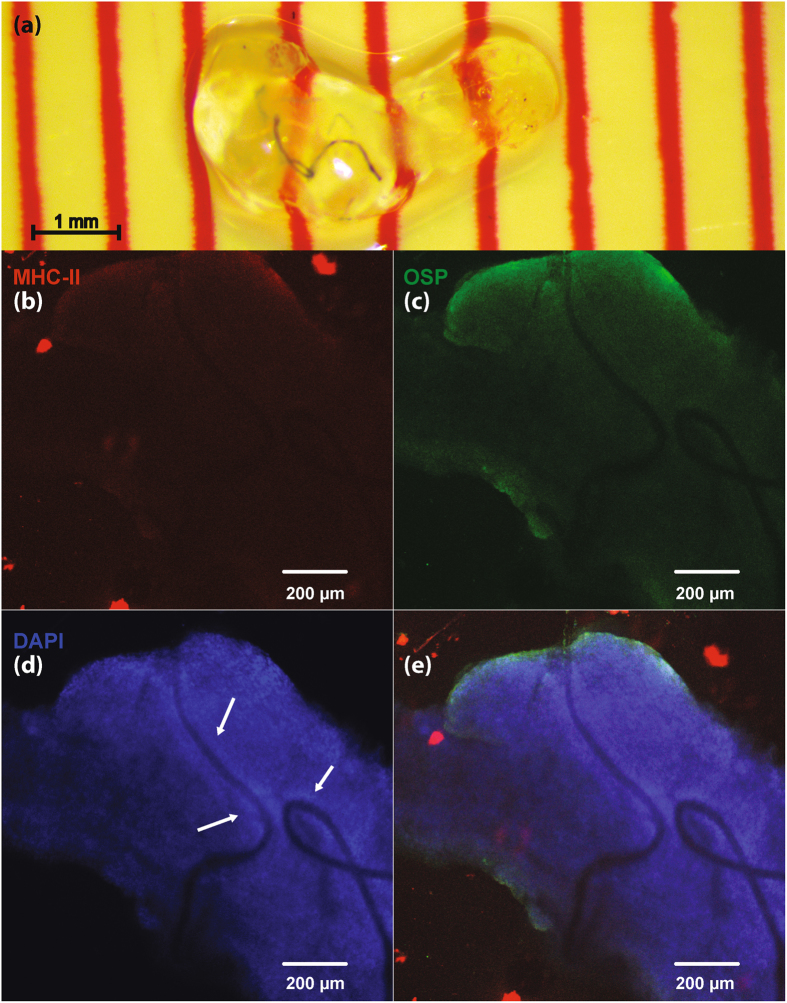
Immunohistology for a chronically implanted VN. Fluorescent images shown at 10x magnification. (a) Cleared VN with implanted CNT yarn electrodes. Nerve shown after 21 days of passive clearing. (b) Fluorescent stain for major histocompatibility complex class II (MHC-II, ab55152), present on antigen-presenting immune cells (e.g. activated microglia). (c) Fluorescent stain for oligodendrocyte specific protein (OSP, ab7474). (d) Fluorescent stain for DNA (DAPI). DAPI is used to stain cell nucleii. Arrows show elevated DAPI density around the wires, used to estimate thickness of fibrous encapsulation. (e) Composite image showing MHC-II, OSP, and DAPI stains.

