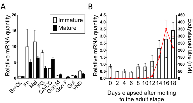
Figure 2.
Tissue and temporal distribution of SgVKR. (A) Tissue distribution of SgVKR in immature (white) and mature (black) adult locusts. Relative transcript levels of SgVKR were measured in different adult tissues using qRT-PCR. All tissues, except the male reproductive system (Gon M), were dissected from immature, 3-day-old female locusts or mature female locusts with an oocyte size between 3 and 6 mm. The male reproductive system was dissected from immature, 3-day-old and mature, 12-day-old male locusts. The data represent mean ± S.E.M. of three independent pools of five animals, run in duplicate and normalized to α-tubulin1A, CG13220 and ubiquitin transcript levels. Abbreviations X-axis: Br + OL: brain + optic lobes; Fb: fat body; Mal: Malpighian tubules; PG: prothoracic glands; CA + CC: corpora allata + corpora cardiaca; Gon M: male reproductive system (testes + accessory glands); Gon F: female gonads (ovaries); Gut: for-, mid- and hindgut; VNC: ventral nerve cord. (B) Temporal distribution profile of SgVKR in the female gonads during the first reproductive cycle. Using qRT-PCR, relative transcript levels of SgVKR were measured every other day in the female gonads, starting on the day of molting to the adult stage (day 0). Data represent mean ± S.E.M. of three independent pools of six animals, run in duplicate and normalized to CG13220 and ubiquitin transcript levels. The ecdysteroid titre (red line), expressed in nM, throughout the first reproductive cycle was measured with an EIA. The data represent mean ± S.E.M. of 6-18 hemolymph samples taken from different animals.

