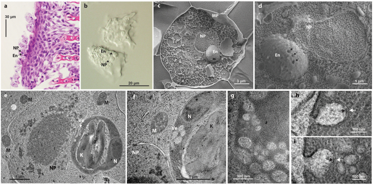
Figure 1.
Paramoeba and its kinetoplastid endosymbiont Perkinsela. (a) Paramoeba sp. cells stained with haematoxylin and eosin in histological sections of gill tissue of Salmo salar (NP = nucleus of the host amoeba; En = Perkinsela sp. endosymbiont). (b) Trophozoites of P. pemaquidensis in hanging drop preparations under Nomarski differential interference contrast microscopy. (c) High-pressure freezing scanning electron microscopy (SEM) of a P. pemaquidensis cell with prominent endosymbiont (MP = plasma membrane of P. pemaquidensis). (d) SEM of the host amoeba nucleus and associated endosymbiont with surface invaginations (arrows). (e–i). Transmission electron microscopy (TEM) of P. pemaquidensis and Perkinsela sp. (e and f) TEMs showing close association of the P. pemaquidensis nucleus (NP) and the endosymbiont Perkinsela sp., the kinetoplast (K) of the endosymbiont, the endosymbiont nucleus (N), vesicles within the endosymbiont cytoplasm (Ve), and mitochondria (M) within P. pemaquidensis. (g–i) TEMs showing ultrastructure of plasma membrane-associated putative endocytotic vesicles within the cytoplasm of Perkinsela sp. White arrows indicate the vesicle membrane, black arrows highlight glycoprotein-rich material on the inner surface of the vesicle, which is continuous with the outer surface of the plasma membrane.

